


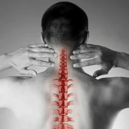
Cervical osteochondria or vertebration occurs as a result of changes in the shape and structure of the vertebrae.Although the cervical area is quite short compared to the total length of the spine, it is perhaps the most important part of the spine.Each pair of adjacent vertebrae forms the intervertebral holes through which nerve roots go and go to each muscle.Through other holes - in the side processes of these vertebrae - vital vessels ensure blood supply to the brain.
The causes of cervical spine osteochondicity
The causes of osteochondry are:
- injuries,
- "Said work" on the screen below eye level,
- Physical work associated with weight transfer,
- Long -term stay driving a car,
- "On the phone" without the use of distance devices (in this case the operator presses the phone on the shoulder of the ear)
- Constitutional Characteristics (Crooked, Claumal Changes in Cervical Vendradims, Short Neck)
Formation of pathological vertebral changes
With osteochondria, the small spots begin to form at the edges of the vertebral bodies, which can injure the structures nearby.Most of the time, this is in response to an excessive load in the cervical compartment and not only is the result of the "aging" of the intervertebral joints (we remind you that it was used to be considered degenerative osteochondria, then a natural age -related illness, such as osteoartica).As the disease progresses, the vertebrae closure plates and the decrease in the height of the intervertebral discs appear.These discs are normal play the role of shock absorption between the vertebrae and, among other things, prevent damage to the spine roots.With the progressive osteochondria, there is a protruding (hernia) of the core of the intervertebral disc jacket, in which there is increasingly pressure during the disease, while weakening the "limitations" of the joints on all sides.This hernia is also capable of compressing vertebral structures and causing neurological manifestations of the disease.
What are the symptoms of cervical osteochondry?
Cervical spine osteochondria
Any pain in the throat causes the pathology of the cervical spine.In terms of growth, the intensity of the pain syndrome is divided into 4 stages, the first patient feels numbness, tingling, "tightening" in the area of a particular muscle group, in the fourth stage - the most severe - the pain is so intense that they lead to its immaculate.
In addition to pain syndrome in the cervical and occipital region, the patient notes "reflecting" (radiating) pain in the upper extremity, the side areas of the chest.
Cervical spine osteochondria
They talk about participating in the process of nerve roots when the pain, numbness and tingling spread to the lower jaw, upper back, forearm and fingers.At the same time, the patient draws attention to the fact that he "seemed to leave" his hand, he was sleeping annoying.Morning stiffness in finger joints, which lasts no more than 10-15 minutes, is noted.With the development of radical syndromes, during the examination, there may be a decrease in the muscle strength of the upper extremities.
Cervical spine bone with "Spine Syndrome"
About participation in the blood vessel process (compressing them with protrusion or osteophytes), they say when the patient complains about frequent attacks, especially after a long stay in a specific position, when he is launched by his head (for example, when he swims with brass)Doppler ”).By ultrasound, the conduct of the spinal arteries, the stenosis of their lumen is determined.In this case, we can talk about surgery, as a strong change in blood flow to spine is a risk factor for the development of the stroke.
Cervical spine bone with "heart (heart) syndrome"
This syndrome forces the patient to come into contact with the cardiologist mainly, as the main complaints are related to pain in the left half of the breast, the sub -tract area, which weaken or intensify when performing physical activity or body position.After excluding myocardial infarction and other heart disease, the patient falls under the observation and treatment of a neurologist and orthopedic.
Diagnosis
To clarify the diagnosis, four methods are used: x -rays, ultrasound, computed tomography and magnetic resonance imaging.
The most affordable method is still the x -ray of the cervical spine, the most informative is the radiography on the side projection ("lateral appearance").This method allows the first approach to determine the presence of injury, gross structural changes in the vertebrae.
Ultrasound examination (ultrasound) is conducted to clarify the condition of the spine.With the help of this method, they discover whether the blood flow is disturbed and if so, to what extent and what kind of obstacles have emerged and where they are identified.
Computed tomography (CT).It allows you to more accurately evaluate the state of bone structures, the degree of bone density, allows you to see smaller osteophytics (bone outgrowths) than possible with x -ray.
Magnetic resonance imaging (MRI).This type of examination is essential for suspected projectile herbs, precise detection of spinal cord damage and the degree of this damage.This study is essential if the question is raised for surgical (surgical) treatment of cervical spine diseases.
Treatment of osteochondry
Medication
The standard set of products for the treatment of cervical osteochondrication reflects the purpose of the treatment: Relieve pain syndrome by removing painful muscle spasm and inflammation of nerve roots, increasing spine motility.To achieve these goals, especially the use of painkillers, nsaids -neculus -scent anti -inflammatory drugs, muscle relaxants are used.It should be remembered that self -interception by these groups can be dangerous, as there is a possibility of incorrect interpretation of symptoms, as well as underestimating the side effects of these drugs.Local (kingdom) drugs between NSAIDs in the form of gel are widely used and when painful pain is, the same drugs can already be used in the form of ointment.
For the treatment of osteochondry in a deeper, "basic" level, preparations are used by a group of chondropr protects containing glucosamine sulfate and chondroitin sulfate.These substances restore the structures of the cartilage of the vertebrae, prevent their further damage.The treatment courses are long, the result remains for many months.
The cervical osteochondria has significant differences from the pathology of the other spine.The pain in the throat in this case may not be caused by signals from the suffering of the spine, but by the painful chronic muscle.It is a completely "benign" condition, which is well treated with the same set of drugs: non -hypotheoid anti -inflammatory drugs with muscle relaxing, using intramuscular "exclusion" using steroids.Usually, the doctor reveals a severe pain when examining SO -called "trigger" spots along the entire cervical spine, as well as in the upper shoulder muscles.Most often such a pathology occurs in women, mainly under 40 years of age.Despite the severe pain syndrome, vascular structures remain intact, the blood flow of the head area does not suffer.
Manual treatment
This method of non -changing treatment may be effective for recently has occurred (often as a result of a minor injury, underground) throat, which is not accompanied by dizziness, other changes from the nervous system and the circulatory system.It is permitted to resort to manual treatment only after a thorough examination, in addition, the doctor performing this procedure should have sufficient experience in the traumatic and orthopedics sector.With the "old" forms of the disease, the use of manual treatment is dangerous!Two methods of this type of intervention are known:
- manipulation (sharp brief influences of significant force aimed at eliminating hypoglycaemia, well -known "bone clicks").
- The mobilization (the method is based on a smooth stretching of the throat after heating and loosening the throat muscles).
It is also used a combined method based in a combination of two mains.It is important to remember that in addition to these contraindications, manual therapy is prohibited for any disease, accompanied by an increase in blood pressure, for any thyroid pathology and ENT or ENT-ORGAN.
Treatment of cervical osteochondicity at home
Medical gym for cervical osteochondria
The first and main rule for beginners to participate in physiotherapy exercises is not to perform exercises, overcoming the painful senses.Of course, you should not start in the "acute" period when the pain just appeared.Another important recommendation is to avoid sudden movements and circular movements in the cervical area.
Each lesson must start with a brief self -sparkling of the muscles of the throat.The following is a "hot" hot -up:
- The hands are reduced, along the body, the shoulders are uniform, the back is straight (you can control the posture with slightly pressed heels, shoulders and buttocks on the wall).Walk in place 1 minute throughout the foot, 1 minute - in socks, 1 minute - on heels.
- The starting position is the same.We break the brushes in fists, reducing our shoulders, our hands are straightened.The moves are slow, we make 20 repetitions, the last rise is more than 5 seconds.We make sure that the throat muscles are not "firm".
- The starting position is the same.We turn our heads in turn to the right, then on the left side.The movements are smooth, a slope in 8 accounts, at the extreme point of the slope - hold for 8 seconds.
- The starting position is the same or sits on a hard chair.Smooth slopes of head forward, at the extreme point - hold for 8 seconds
- The starting position is the same or sits on a hard chair.Slowly sloping head forward, until the chin on the chest, then slowly turn your head to the right (in 4 accounts) and to the left (in 4 accounts).Do not allow muscle strain.
- The starting position is the same or sits on a hard chair.We increase your shoulders to 4 accounts, then we also reduce them to 4 measurements.10 repetitions.
- The starting position is the same or sits on a hard chair.We are increasing our shoulders, but now we are running circular movements back, 8 accounts.10 repetitions.
- Align the back, check the posture (see exercise 1).In 4 accounts, we reduce the shoulders behind your back, trying to connect them, to the end point for 8 seconds, then return to the starting position.
Pillows
As already mentioned, the hypertension of the throat muscles is the first and often the main reason for the growth and evolution of cervical osteochondrication.A rational choice of pillows and layers, ensuring that a relaxed and comfortable position during sleep is no less than fitness, naturalness and drugs.
When selecting a mattress, pay attention to the filler composition (the products are suitable, at least half of coconut chips, that is, with sufficient rigidity).Soft layers of spring do not provide adequate alignment of the spine.The most optimal sleep to sleep is on the side, pull one or both knees in the stomach.The pillow must be in such a way that it fills the whole space between the shoulder, ear and matt, the head (crown) of the head is in the same horizontal line with the spine.To avoid too high and very low, as well as soft pillows.The ideal choice is a polyurethane foam of an ergonomic shape, that is, in this case, with a slight compression on one side.
General recommendations
Pay attention to posture.During walking or at a stand, the position is a position when the chest protrudes forward and the stomach is pulled.
Avoid long -term stay in a sitting position.A simple rule of prevention of cervical osteochondrication is well known: after every 60 minutes of work, a 10-15-minute hiking period is required.
A chair for work should have a high headrest or back.
In a sitting position, the legs must be based on the floor and the neck should not be tense.For this purpose, use special orthopedic devices: cylinders under the neck while driving in a car, a pillow under the back.
Avoid lifting weights.If necessary, kneel, press the heavy object on the body and then stood smoothly using the strength of the leg muscles, but not the "push" of the back.
Do not rest with straight legs.Use kiosks, work surfaces to bring the object closer to yourself and not persuade your face on the subject.Try to do homework at home in a chair or a fitness ball.
If you need to use a mop, broom or rake, do not stretch your hands, back and neck, do not rest sideways.
Avoid swimming in brass style.